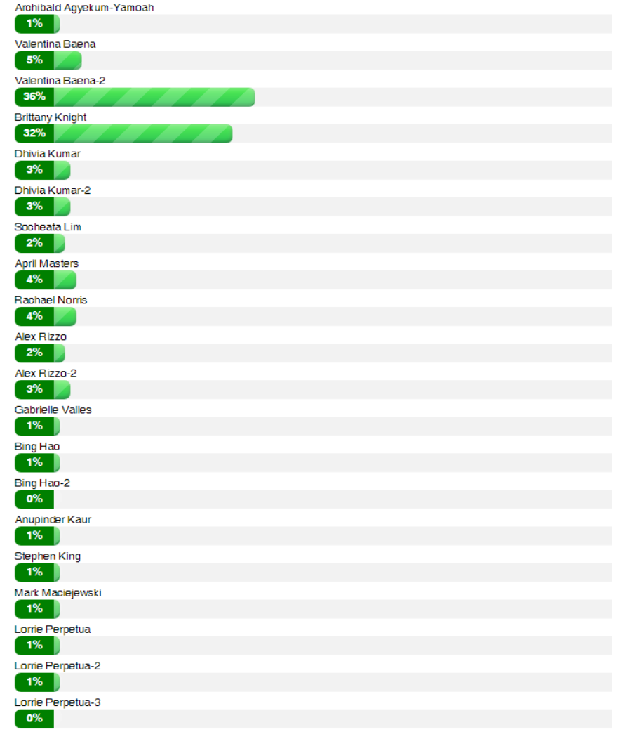
Contest Winners:

Archibald Agyekum-Yamoah
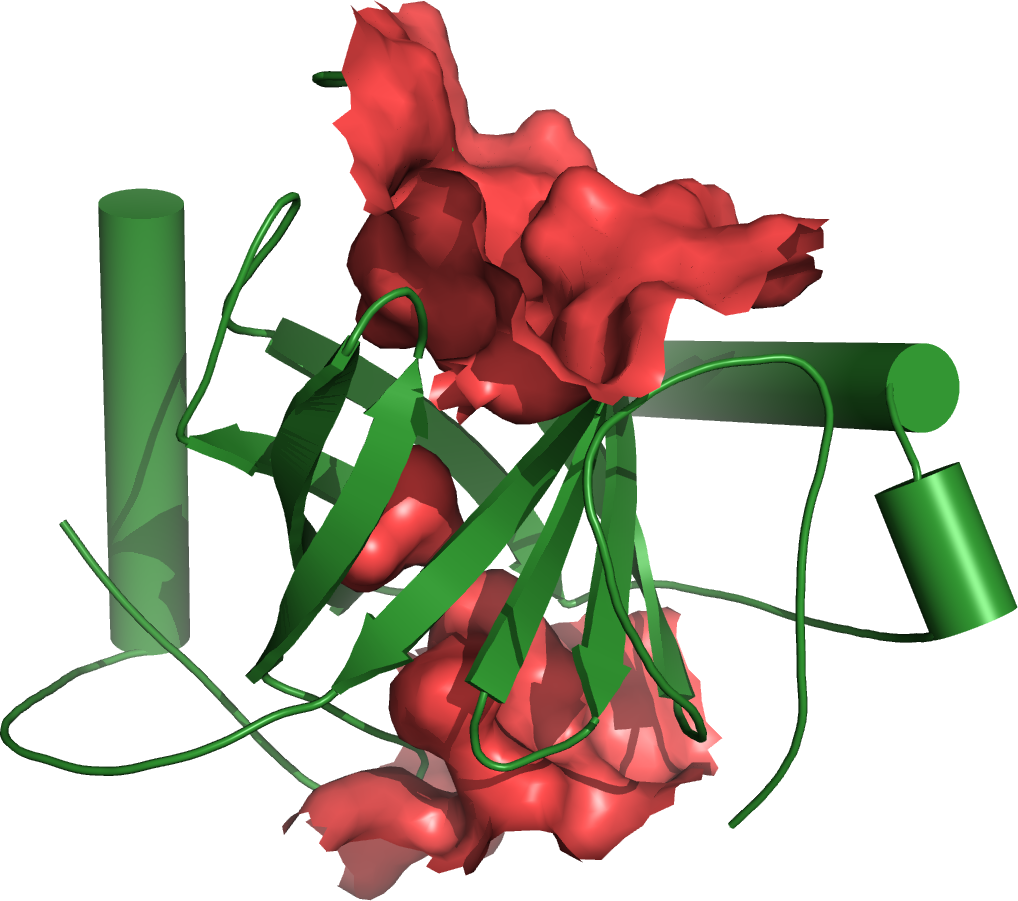
Valentina Baena

Valentina Baena-2
Brittany Knight
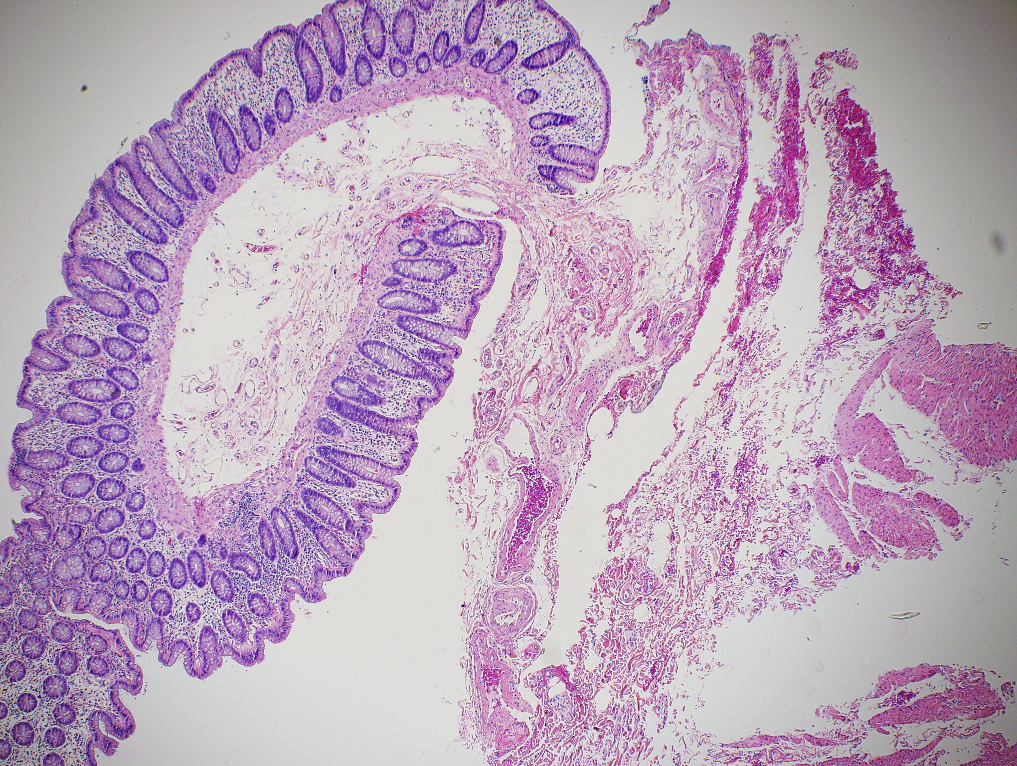
Dhivya Kumar
Dhivya Kumar-2
Socheata Lim
April Masters
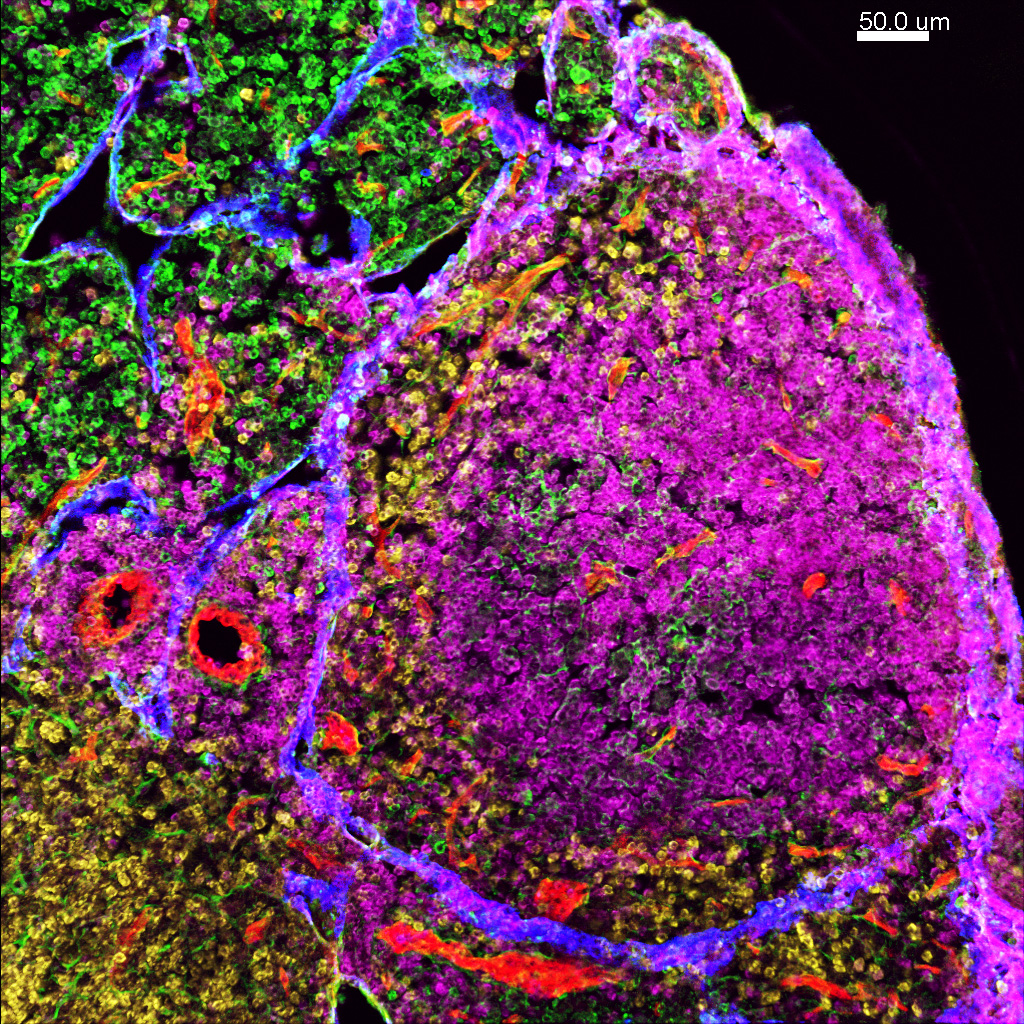
infection.
Cell- Maker- Color
B cells-B220 -pink
Stromal cells- Podoplanin-green
T cells -CD8a -yellow
Blood vessles-CD31- red
Lymphatic vessles- Lyve-1- blue
Rachael Norris
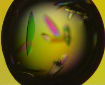
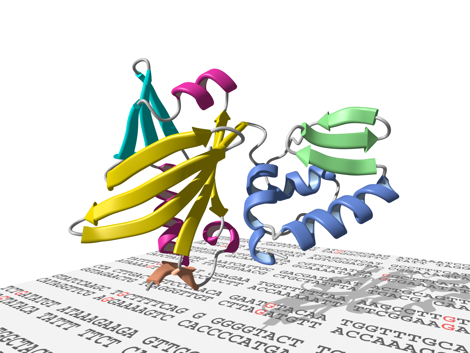
Alex Rizzo
Alex Rizzo-2
Gabrielle Valles
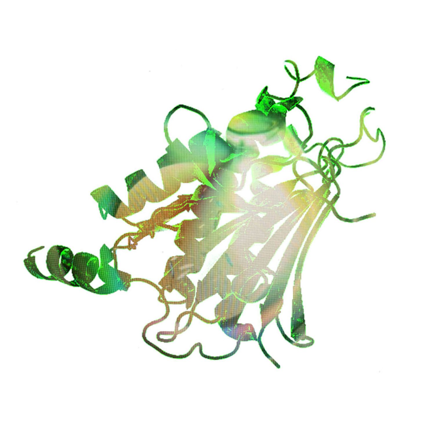
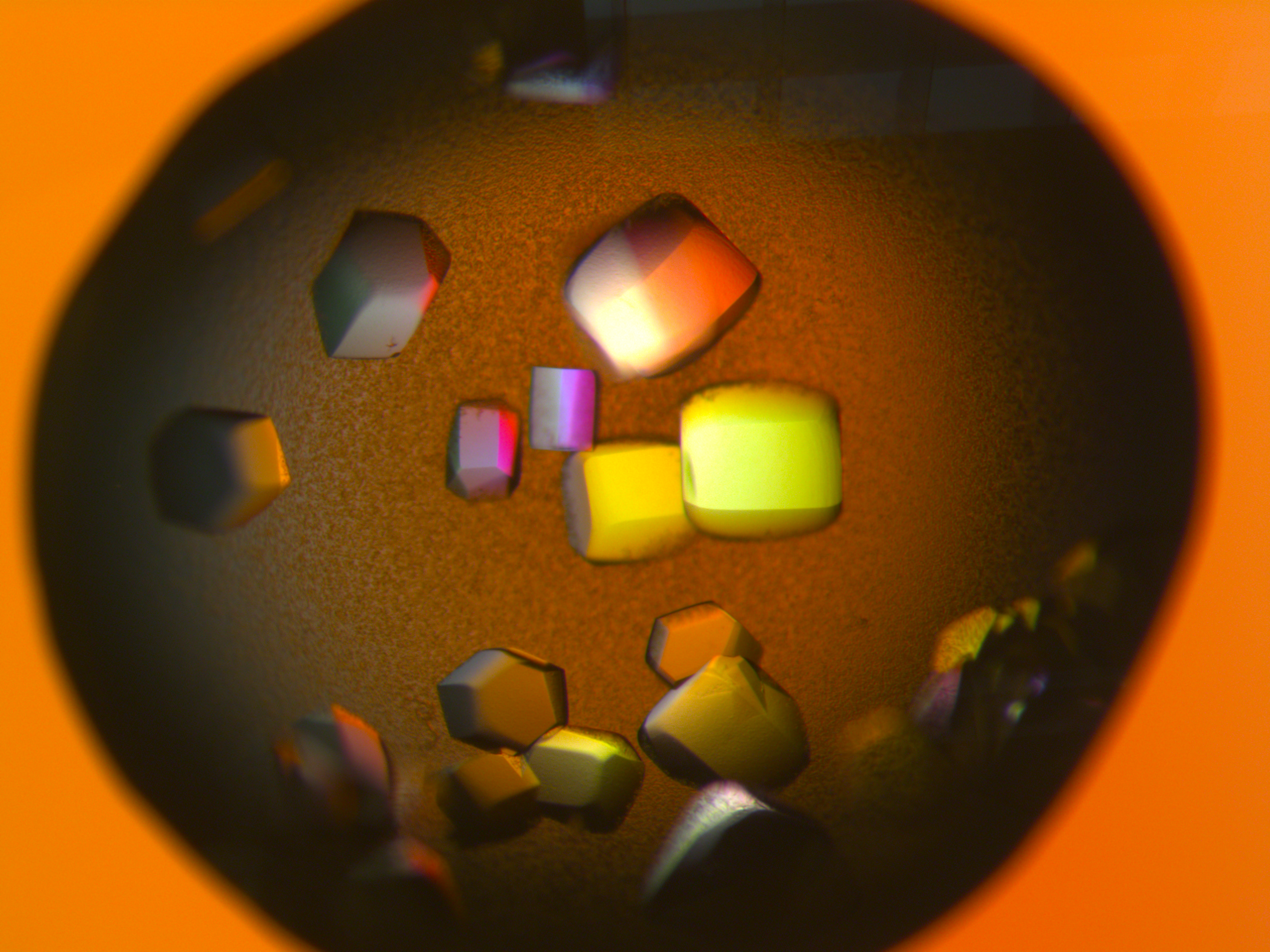
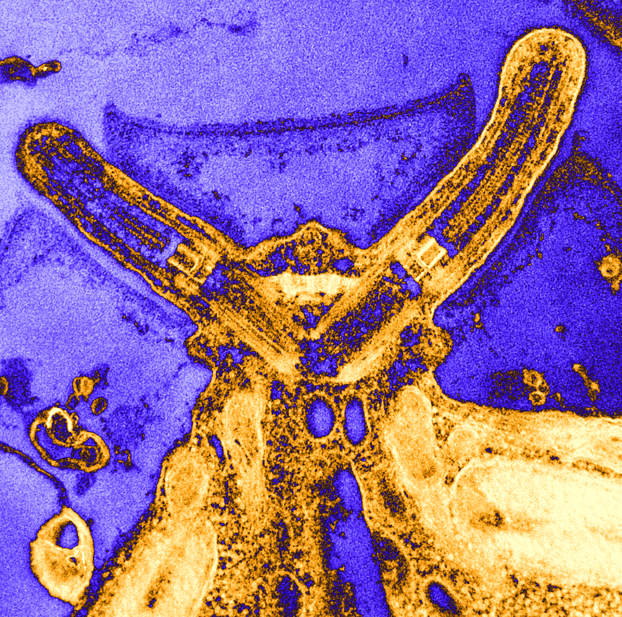
Bing Hao
Bing Hao-2

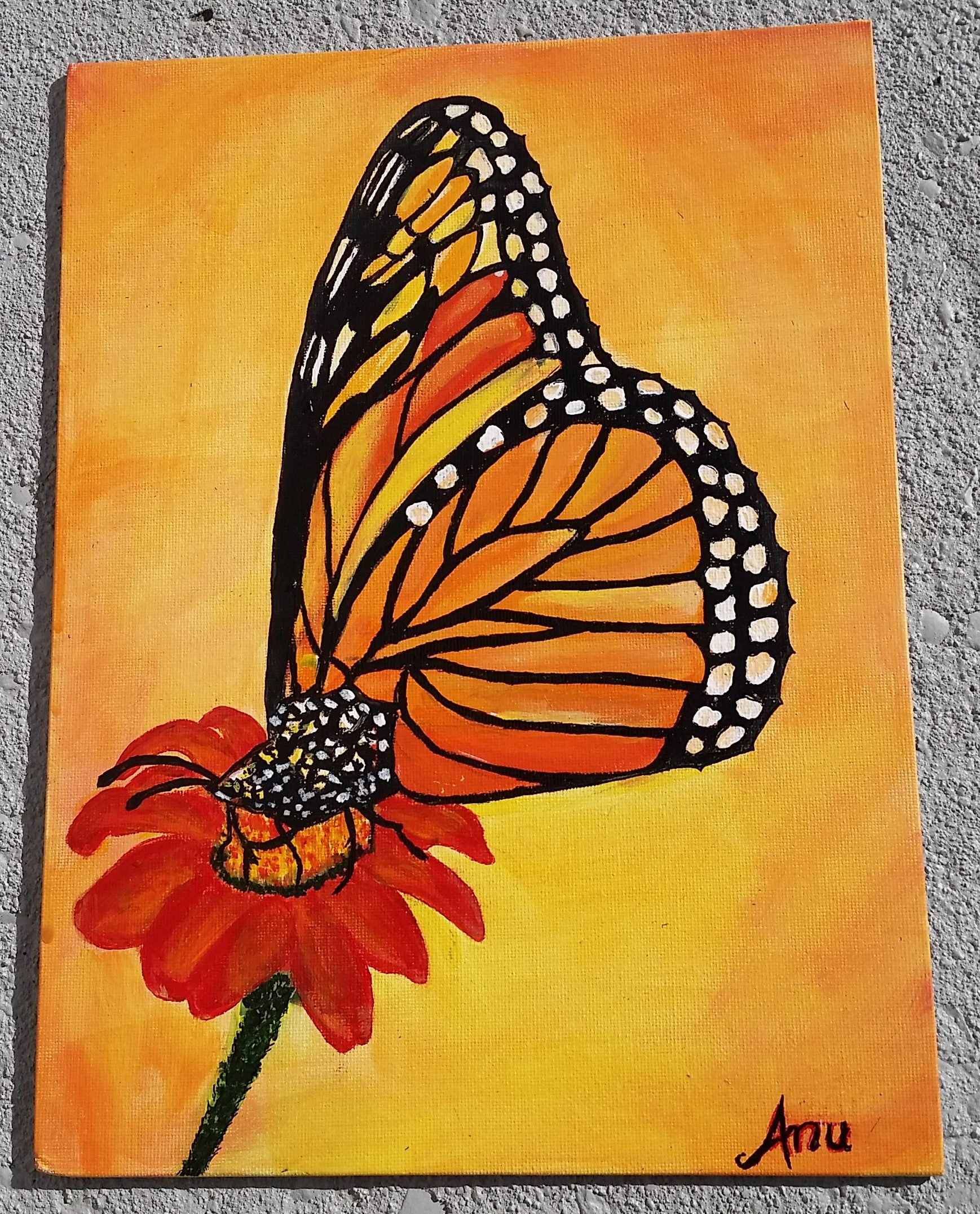
Anupinder Kaur
Stephen King
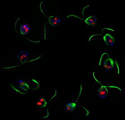
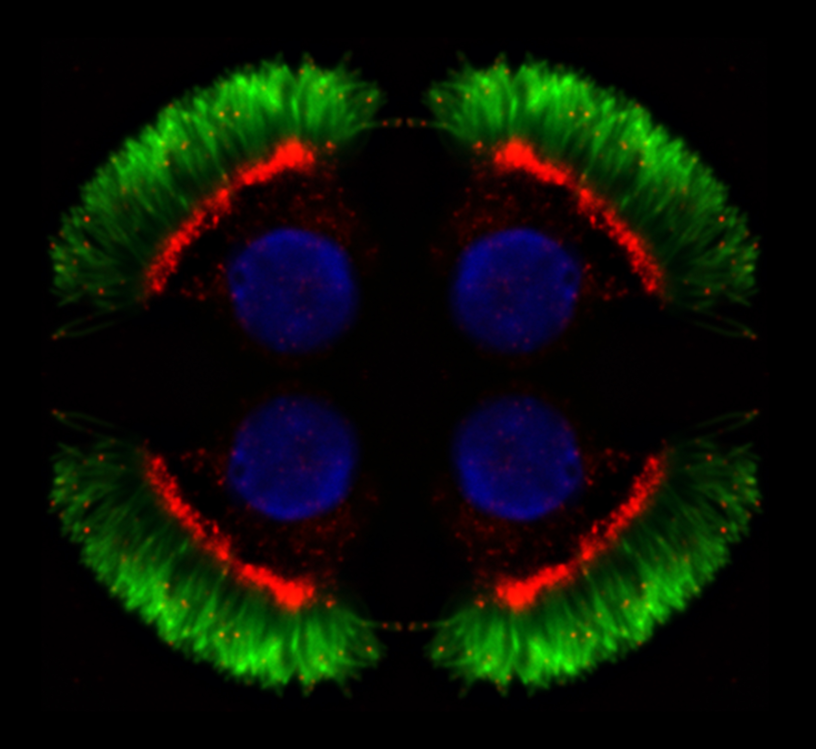
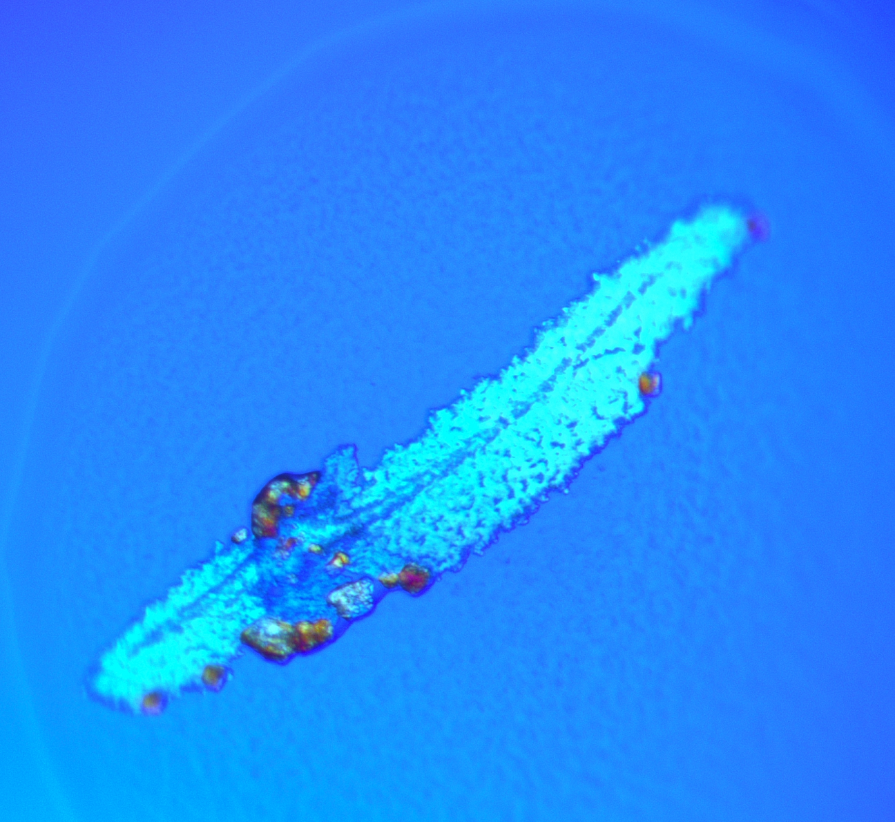
 Short cilia of a mutant Chlamydomonas.
Mark Maciejewski
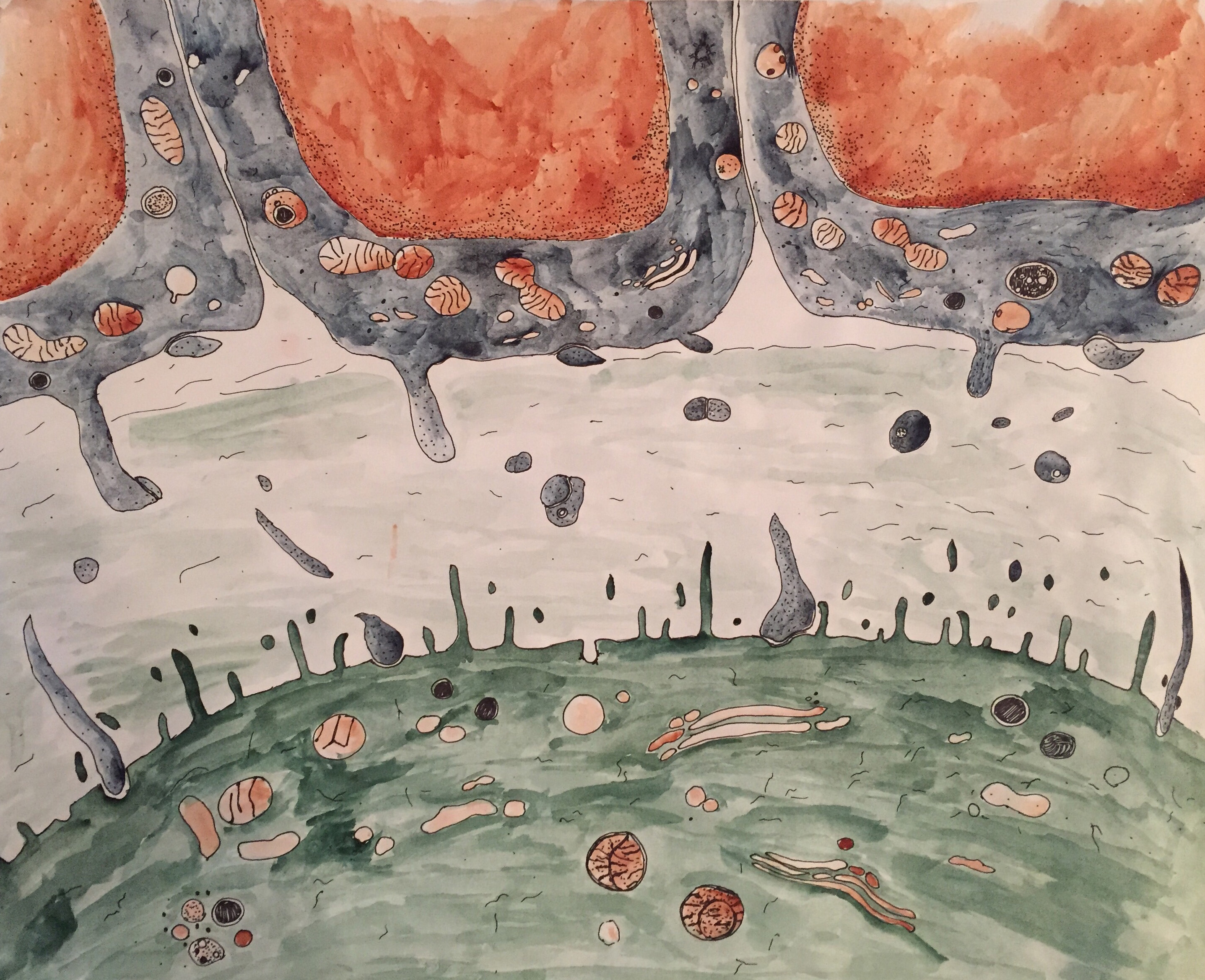
Lorrie Perpetua
Lorrie Perpetua-2
Lorrie Perpetua-3
poll-results