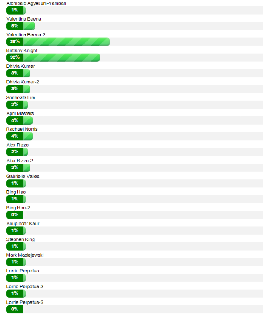
Contest Winners:

Archibald Agyekum-Yamoah
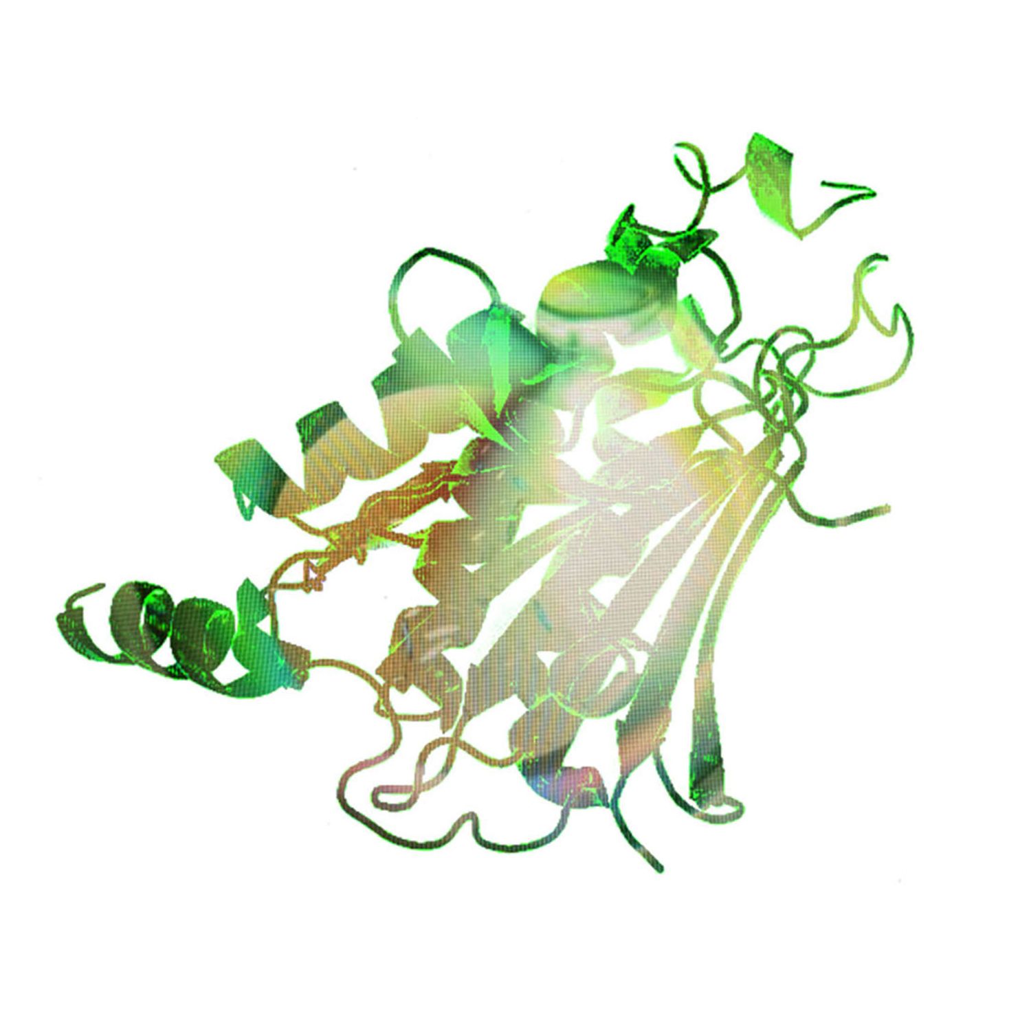
Valentina Baena

Valentina Baena-2
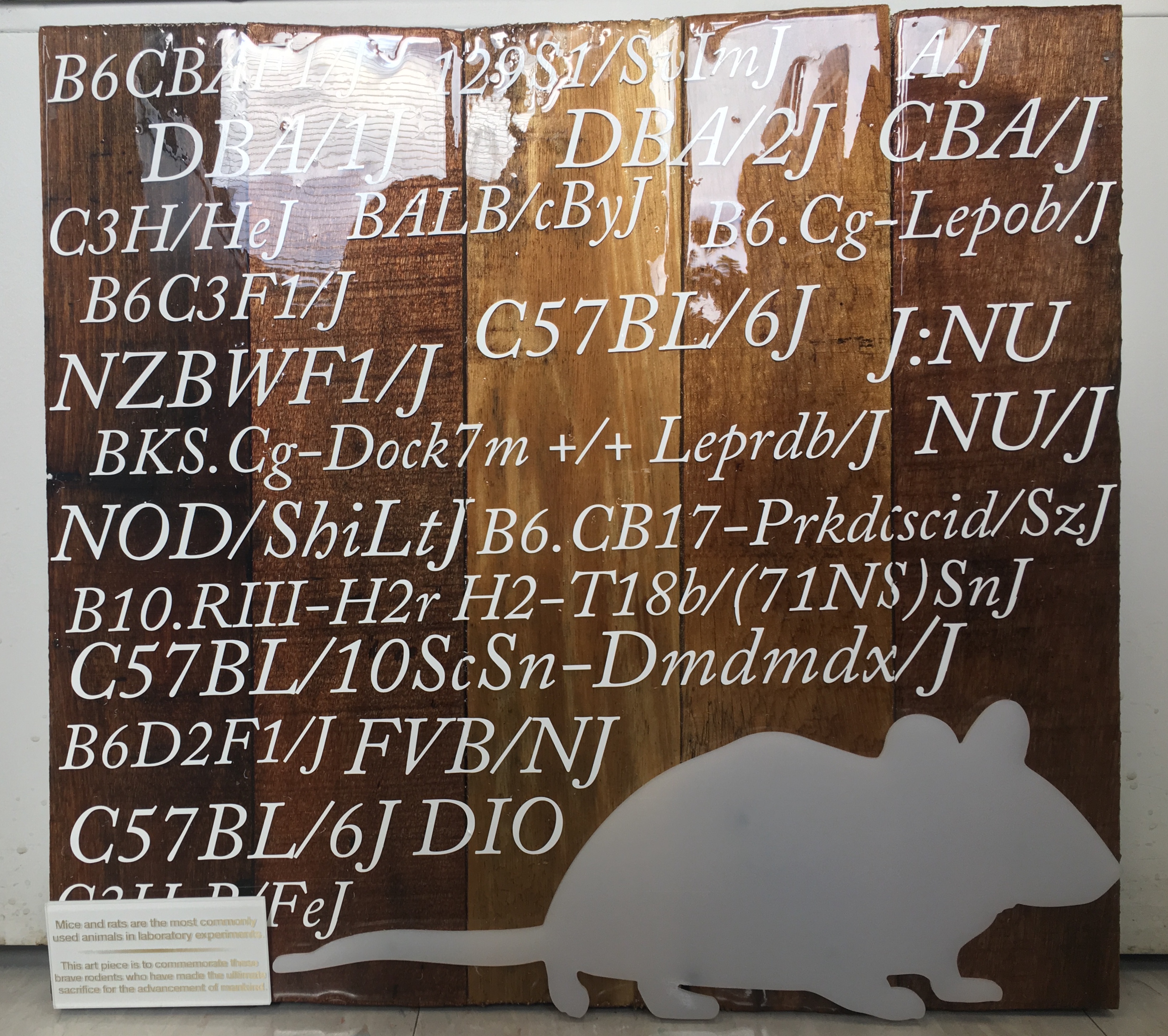
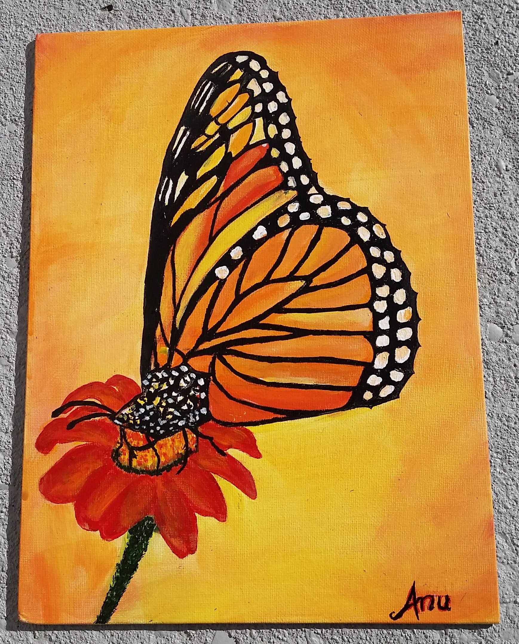
Brittany Knight
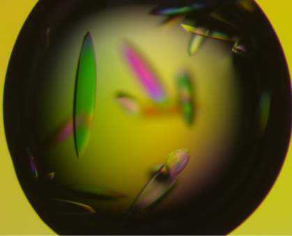
Dhivya Kumar
Dhivya Kumar-2
Socheata Lim
April Masters
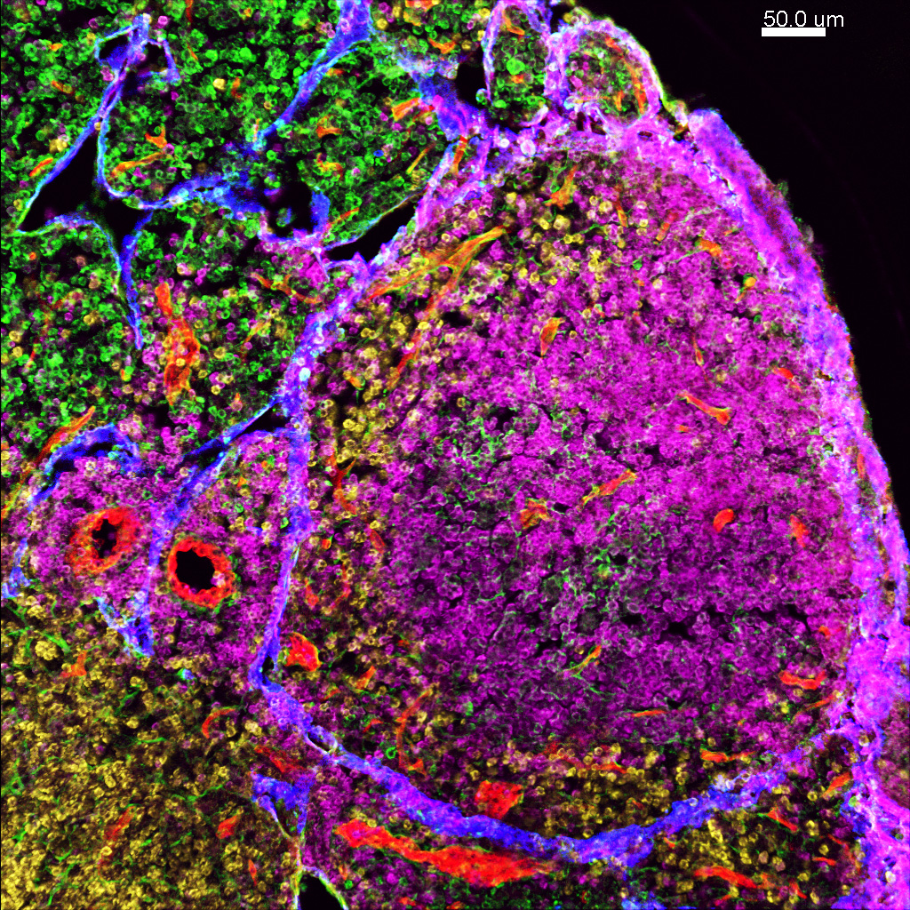
infection.
Cell- Maker- Color
B cells-B220 -pink
Stromal cells- Podoplanin-green
T cells -CD8a -yellow
Blood vessles-CD31- red
Lymphatic vessles- Lyve-1- blue
Rachael Norris
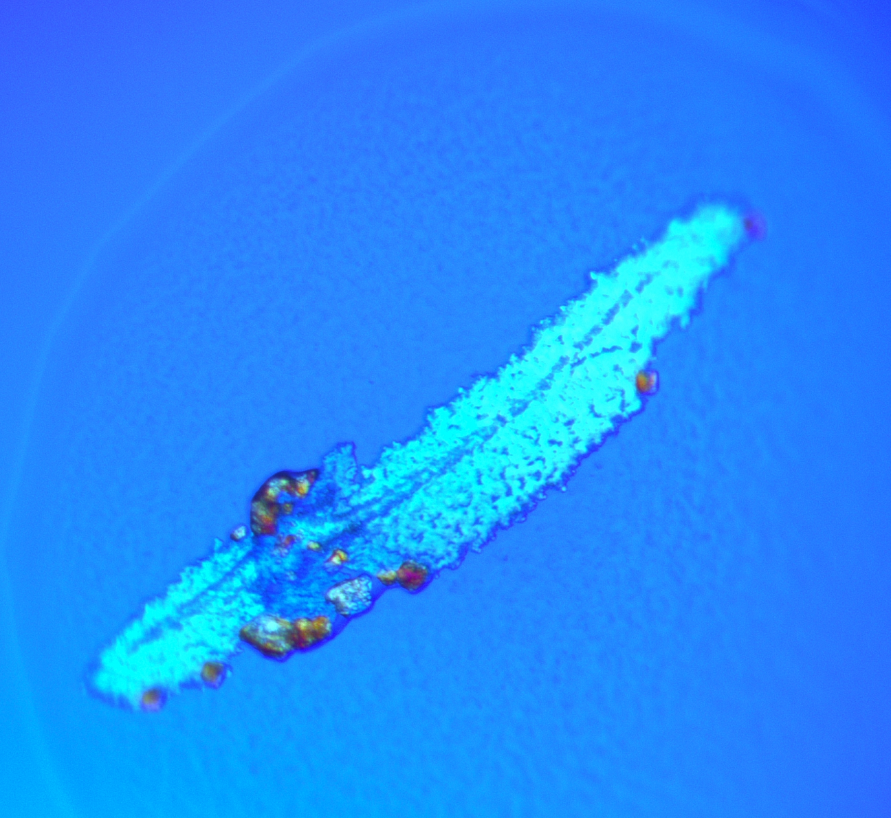
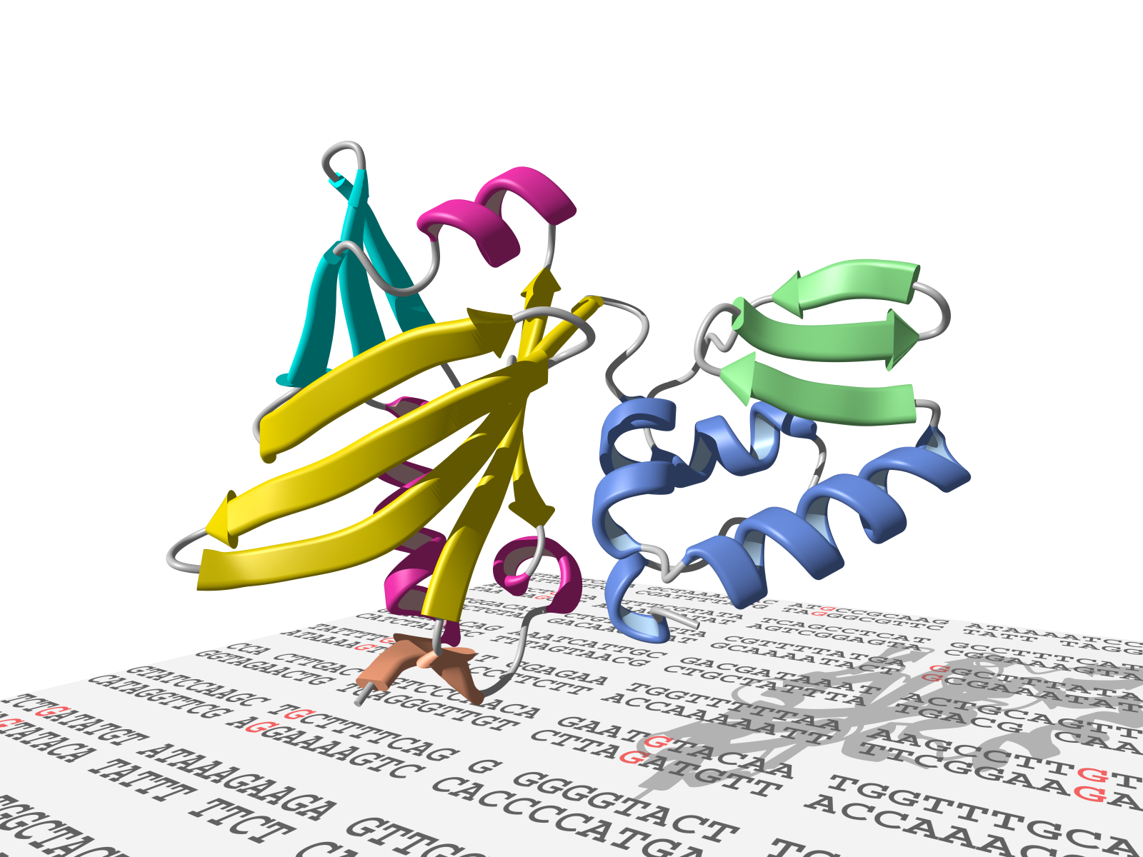
Alex Rizzo
Alex Rizzo-2
Gabrielle Valles
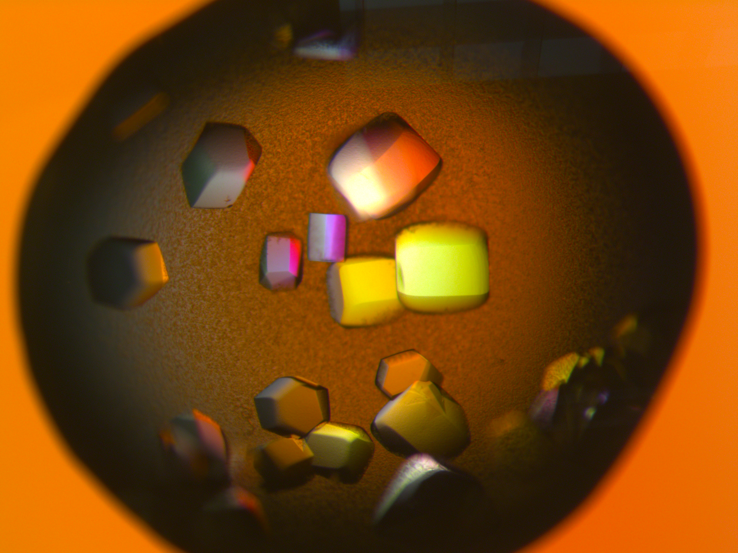
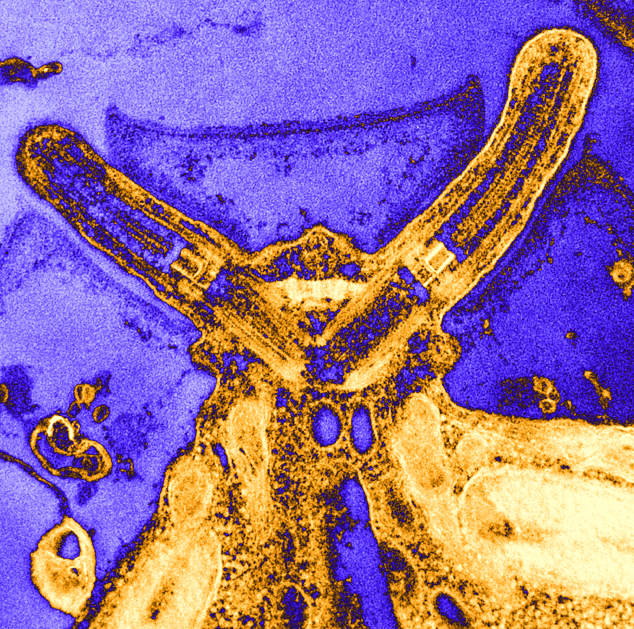
Bing Hao
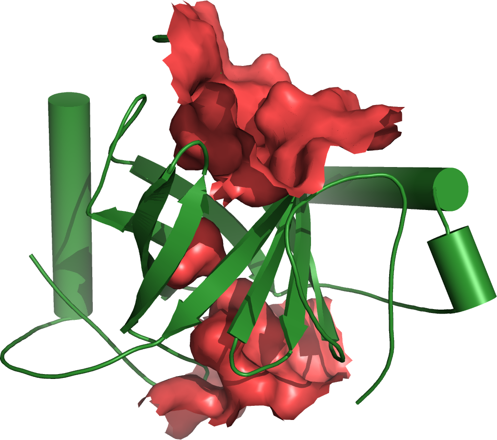
Bing Hao-2

Anupinder Kaur
Stephen King
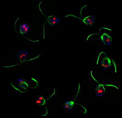
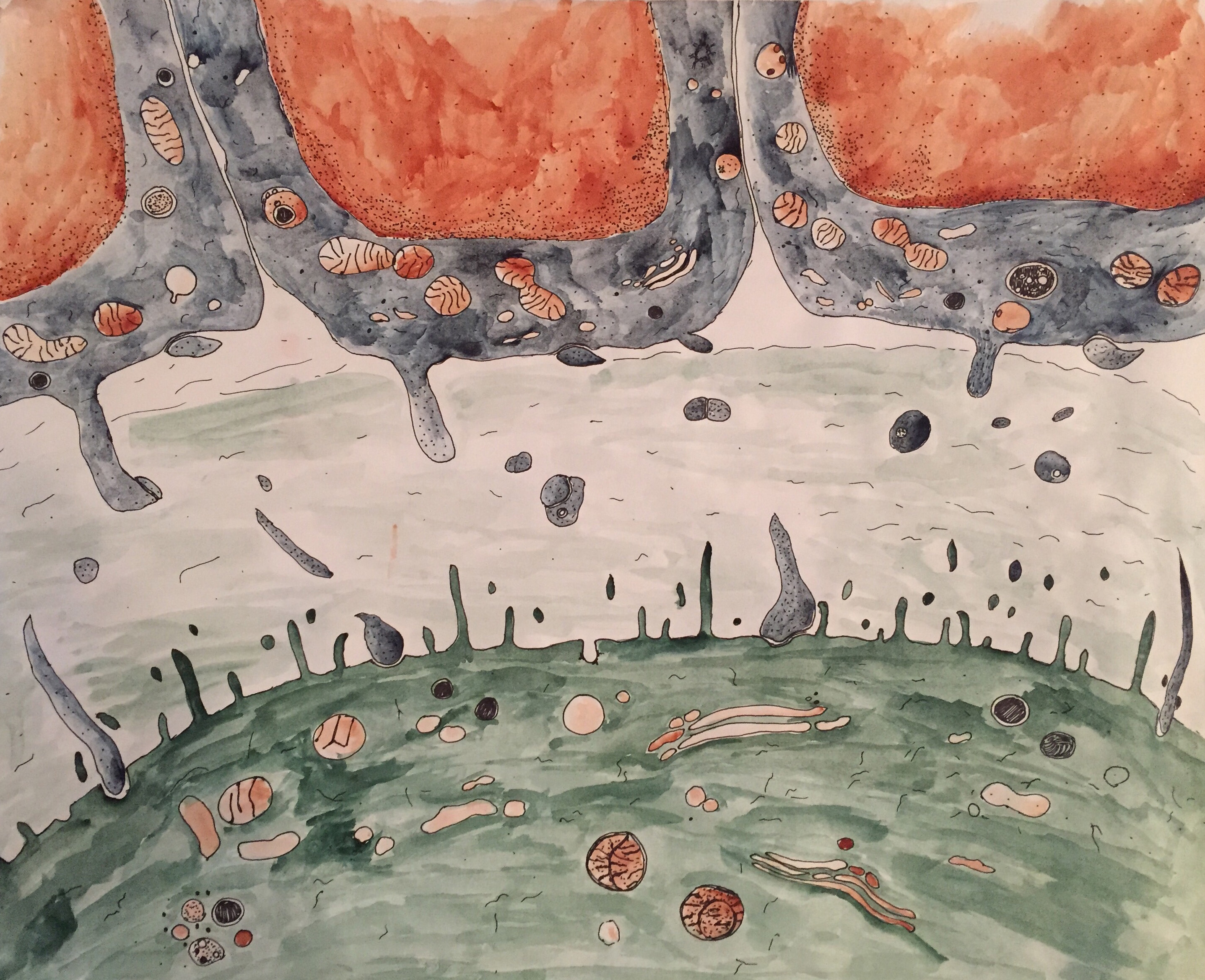
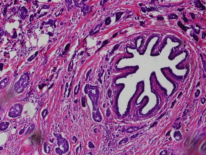
 Short cilia of a mutant Chlamydomonas.
Mark Maciejewski
Lorrie Perpetua
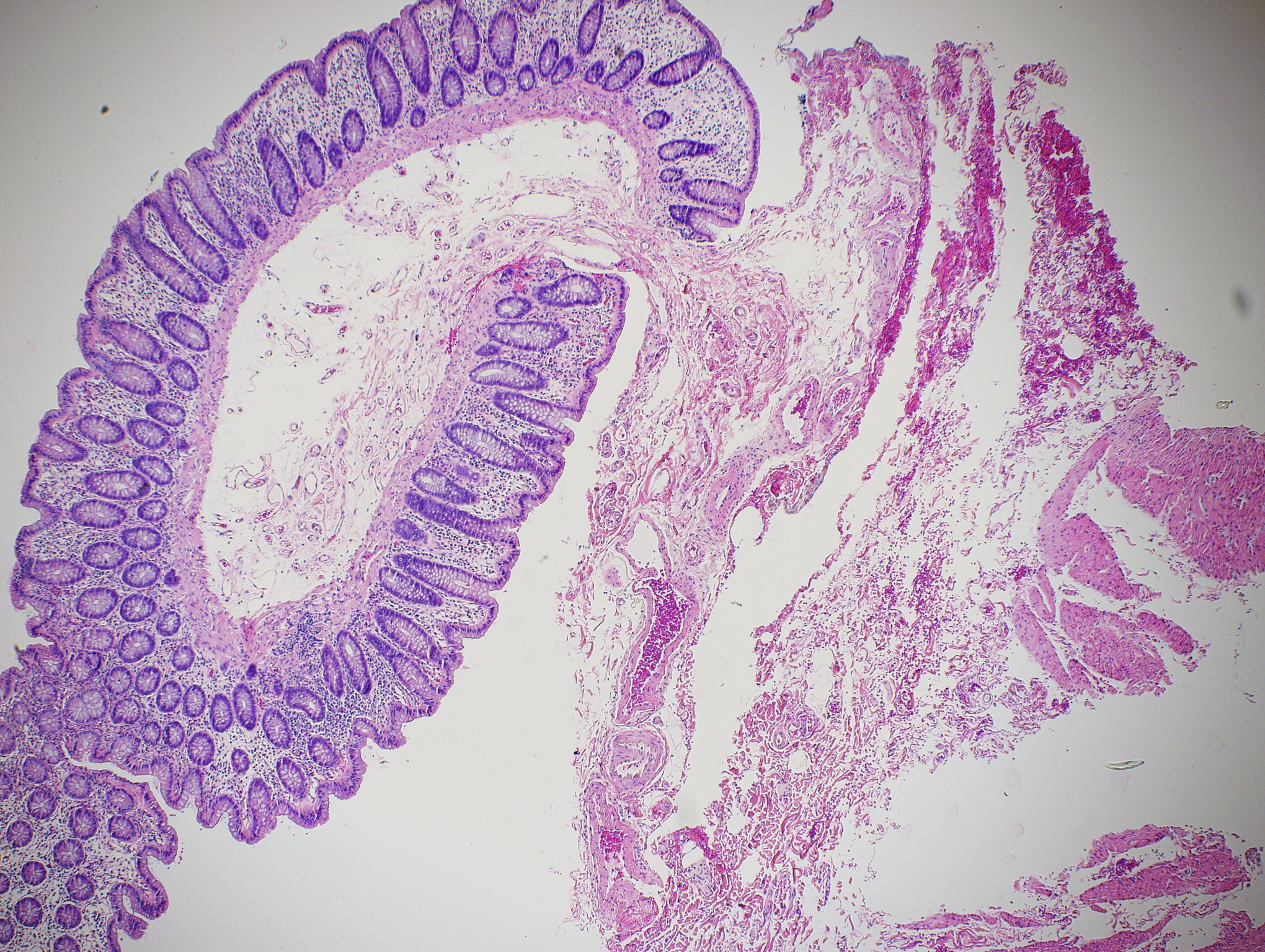
Lorrie Perpetua-2
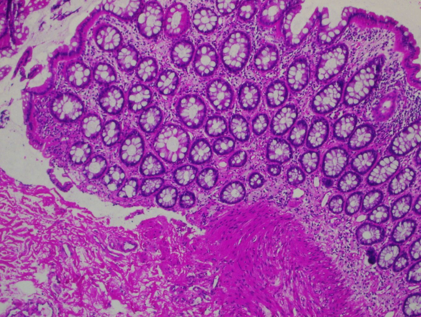
Lorrie Perpetua-3
poll-results
Huge range of beautiful images. Thanks to all!
I agree with Betty. I love so many of them!
There is so much great artwork here. Great job everybody. For me, the true meaning of this art contest was to create an original piece of science inspired art. Those students who went above and beyond with a cartoon rendering of their own results, such as Rachael Norris, and creating something completely unrelated to what they study, but recognizing the animal subjects used in research (Britt Knight) should be recognized.